Dental X-Rays
More On General Oral Care
About General Oral CareFillings
Tooth Decay
Tooth Jewelry
Bad Breath
Bruxism
Sedation Dentistry
There are many diseases of the teeth and surrounding tissues that cannot be seen when your dentist examines your mouth. If X-rays are not used, small cavities between teeth, abscesses, cysts, tumors, and other diseases may be impossible to detect until obvious signs and symptoms have developed and serious damage has been done to your health.
Women must tell their dentist if they are pregnant. When a pregnant woman wears a leaded apron during dental X-rays it is unlikely that the developing baby receives any detectable radiation from outside the body. Today the lead aprons offer more peace of mind than actual protection because stray radiation from modern dental X-ray machines is almost nonexistent.
For adults, radiographs can:
• Show areas of decay that your dentist may not be able to see with just a visual examination, such as tiny pits of decay that might occur between teeth
• Find decay that is developing underneath an existing fillings
• Find cracks or other damage in an existing filling
• Alert the dentist to possible bone loss associated with periodontal (gum) disease.
• Reveal problems in the root canal, such as infection or death of the nerve
• Help your dentist plan, prepare and place tooth implants, orthodontic treatments, dentures or other dental work
• Reveal other abnormalities such as cysts, cancer and changes associated with metabolic and systemic diseases
The different types of X-rays: These are divided into two main categories: intraoral, which means that the X-ray film is inside the mouth; and extraoral, which means that the film is outside the mouth.
Intraoral radiographs:
The various types of intraoral X-rays show different aspects of the teeth:
Bite-wing X-rays highlight the crowns of the teeth. On each radiograph, the upper and lower teeth in one portion of the mouth are shown, from the crown to about the level of the jaw.
Periapical X-rays highlight the entire tooth. On each radiograph, the teeth from either the upper or lower jaw in one portion of the mouth are shown. The difference from bitewings is that in a periapical X-ray, the whole tooth is shown, from the crown down past the end of the root to the part of the jaw where the tooth is anchored.
Periodically, a dentist may recommend a "full-mouth radiographic survey," or FMX. This means that every tooth, from crown to root to supporting structures, will be X-rayed using both bitewing and periapical radiographs.
Occlusal X-rays are larger and highlight tooth development and placement. On each radiograph, nearly the full arch of teeth in either the upper or lower jaw is shown. These X-rays are taken with the X-ray machine either pointing straight down from near the nose (to take pictures of the upper jaw and teeth), or straight up from under the chin (to take pictures of the lower jaw and teeth).
Digital radiographs are one of the newest X-ray techniques around. With digital radiographs, film is replaced with a flat electronic pad or sensor. The X-rays hit the pad the same way they hit the film. But instead of developing the film in a dark room, the image is electronically sent directly to a computer where the image appears on the screen. One of the great advantages of this process is that radiographs can be digitally compared to previous radiographs in a process called subtraction radiography. The computer can digitally compare the two images, subtract out everything that is the same and give a clear image of anything that is different. This means that tiny changes that may not be noticeable with the naked eye can be caught earlier and more clearly with digital-subtraction radiography.
Extraoral radiographs:
Extraoral X-rays are made with the film outside the mouth. Extraoral X-rays are less detailed than intraoral X-rays, so they are not used for detecting caries or flaws in individual teeth. They show teeth, but their main focus is on the jaw or skull. Extraoral radiographs are used for monitoring growth and development, looking at the status of impacted teeth, examining the relationships between teeth and jaws and examining the temporomandibular joint or other bones of the face.
Panoramic radiographs show the entire mouth area - all teeth on both upper and lower jaws - on a single X-ray. This type of X-ray requires a special panoramic X-ray machine. The tube head that emits the X-rays circles behind the patient's head, while the film simultaneously circles across the front. That way, the full, broad view of the jaws is captured on one film. The machines may have chin rests, forehead rests, and side head positioners, plus bite-blocks that patients will be asked to close their teeth around. But the process is very safe and often uses less radiation than intraoral radiographs.
Tomograms are a special type of radiograph in which the dentist can focus in on one particular layer, or slice, of anatomy while blurring out all other layers. This allows dentists to see structures that may be difficult to see with standard X-rays, for example, the temporomandibular joint. The condyle that makes up part of the joint is in the middle of a dense cranial base, so it is extremely difficult to X-ray. Using a tomography technique called a temporomandibular joint projection, a straight “slice" that's lined up with the condyle shows that area more clearly.
Cephalometric projections are X-rays taken of the entire side of the head. They are used to look at the teeth in relation to the jaw and the profile of the individual. Orthodontists use Cephalometric projections to plan their treatments. They will look at the entire face to determine the best way to get the teeth aligned in the right way for that particular person, according to the size of their teeth and jaws.
Sialography is a way of visualizing the salivary glands on a radiograph. Soft tissues, like gums and salivary glands, can't usually be seen on an X-ray because they are not dense enough to absorb enough X-rays to appear clearly on film. With sialography, the dentist injects a radiopaque contrast material directly into the salivary glands. This material shows up easily on film, allowing dentists to diagnose salivary gland problems, such as blockages or Sjögren's disease.
Computed tomography, or CT scanning, usually is performed in a hospital, not the dentist's office, although a dentist may refer a patient for this test. With this process, the patient lies still in the CT machine while the X-ray beam rotates around. From the X-ray information, a computer creates a three-dimensional image of the interior structures. It is used to identify problems in the bones of the face, such as tumors or fractures.
There is a lot of confusion and concern about the radiation from dental radiographs. The diagnostic X-rays affect only a small part of the body for a very short time. Exposure to large amounts of X-radiation is harmful. But with modern techniques and equipment, the amount of radiation received in a dental examination is extremely small. The new digital radiograph systems produce less than 1/5 the radiation that the older film systems use.
The other factor is the type of tissue exposed to radiation. Radiation damage is mostly only a consideration with rapidly dividing tissues such as reproductive organs, bone marrow, etc. The tissues exposed to dental x-rays (teeth, jaw, cheeks) are not as susceptible to radiation damage. Therefore, the risk of harmful effects from dental X-rays is negligible.
It is difficult to determine whether small doses of X-rays increase the individual patient's risk of cancer. However, the chances are extremely small that dental X-rays contribute to cancer, because the exposure of the tissues of the head and neck is so small. Even so, X-ray examinations should be made only when necessary. Many times a radiograph is necessary to diagnose certain conditions; and therefore, the benefit outweighs the risk.
An important advantage to this new digital X-rays technology is that it reduces the amount of radiation by 90% and is much faster. The digital image only takes about 20 seconds to appear in the monitor. In addition, this new technology is friendly to the environment since no chemicals are needed for developing the image.

More dental deals in Other Cities
50 % OFF
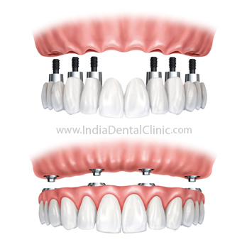
Full Mouth Dental Implant Fixed Teeth
Special Price - 20000 (INR-Indian Rupee )Treatments Covered-
- Dental Implants
- Implant Bridges
- Full Arch Implant Dentures (U&L)
- Full Arch Implant Bridges (U&L)
View Details Nashik
50 % OFF
50% OFF ON IMPLANTS
Special Price - 24999 (INR-Indian Rupee )Treatments Covered-
- Dental Implant Consultation
- Dental Implants
- Dental Sealants
View Details Bengaluru